
Scientists were able to record the moment in real time and create a three-dimensional image of it for the first-time. ViralThe hijacking of a cell gives us a deeper understanding of how infections spread in the body.
The two-and-a half-minute film shows microscopic nature. Genetically sterileAs it seeks an entry point, viruses are thousands of times smaller in size than a grain if sand.
Understanding how Viral infections break into cells is crucial in working out better ways of defending against them, but tracking these particles is incredibly difficult – not least because they are so much smaller than the cells they’re navigating.
“It’s almost like you’re trying take a photograph of someone standing in front a skyscraper.” Courtney Johnson, a chemistDuke University in North Carolina. “One photo is not enough to capture the skyscraper in its entirety and the details of the person who lives there.”
Furthermore, virus particles travel faster outside cells than inside them, making it more challenging to create an imaging process that is sensitive to these differences.
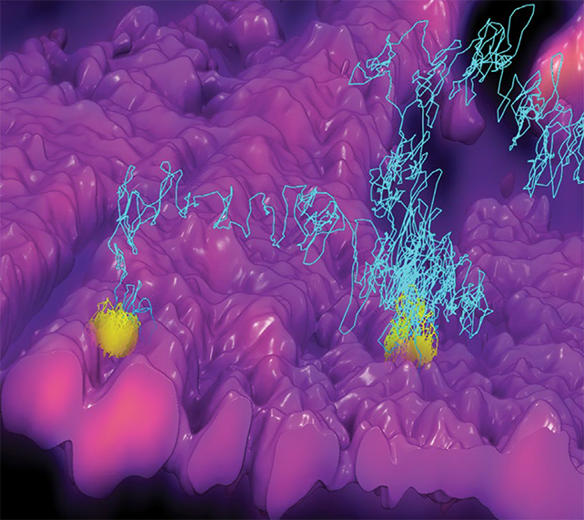
3D-TrIm (or 3D Tracking and Imaging Microscopy) is the solution. It is basically two microscopes in one. The first can ‘lock on’ the fast-moving particles and the second can capture 3D images of surrounding cells. It works in a similar way to a satellite navigation app that tracks your car’s position in the middle of larger landscapes.
The fluorescent label illuminates the virus particle and allows researchers to plot its position 1,000 times per second. Researchers can then see the movement of the virus particle over a crucial period in the infection process with unprecedented detail.
The virus’s meandering path can be seen in the Duke University video below as a purple line.
frameborder=”0″ allow=”accelerometer; autoplay; clipboard-write; encrypted-media; gyroscope; picture-in-picture” allowfullscreen>
“Sometimes, when I present this work, people ask me if it’s a videogame or a simulation.” Johnson. “No, it’s not. This is what came out of a real microscope.”
We’re all breathing in millions of viruses every day, the vast majority of which fail to do any harm – but scientists want to learn more about how certain viruses break through the protective layer of cells and mucus covering the airways and gut to establish an infection.
The 3D-Trim technique should be helpful, but it does have its limitations. Before imaging virus particles, they must be labeled. Additionally, the fluorescent dye used on them must be long lasting enough to allow researchers to track the entire infection process.
However, the team behind 3D-Trim says the potential is there for the system to get better quickly, and to be adapted to other types of medical diagnostics – whether that’s keeping watch over viruses or monitoring drug delivery.
The researchers write in their paper that “Importantly this technique can also be applied to any system where fast dynamic of nanoscale objects occurs over large volumetric sizes, including delivery nanoscale drug candidates into the lungs and through leaked tumor vasculature.” Published paper.
The research was published in Nature Methods.
