
Scientists have just discovered a significant new clue that may help to solve the frustrating mystery of the migraine.
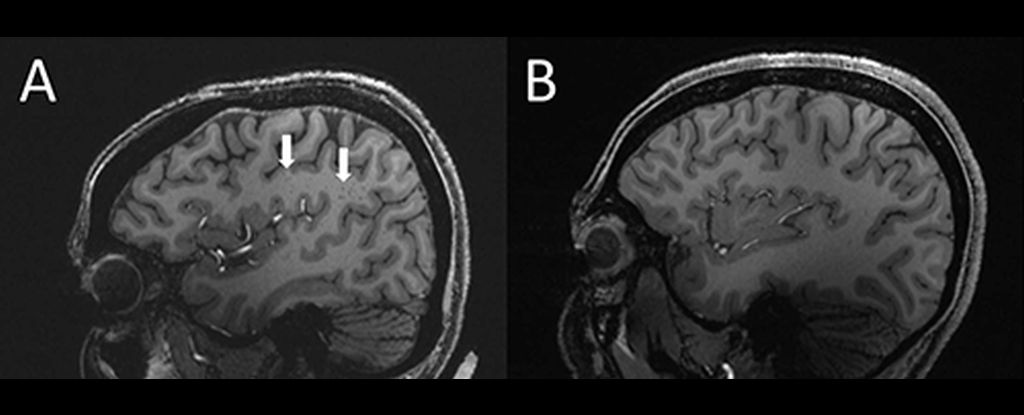
Using ultra-high-resolution MRI, researchers found that perivascular spaces – fluid-filled spaces around the brain’s blood vessels – are unusually enlarged in patients who experience both chronic and episodic migraine.
While the relationship to migraine and its role is still not clear, this finding could be a promising avenue for future research.
The discovery was presented at both the 108th Scientific Assembly, and the Annual Meeting of Radiological Society of North America.
“In people with Chronic migraine and episodic migraine“Without aura, there are significant differences in the perivascular areas of a brain area called the centrum Semiovale. Wilson Xu is a medical scientistLos Angeles University of Southern California
“These changes were not reported before.”
Migraine is a terrible condition. While migraine is most well-known for its severe headaches, it can also be caused by migraine. vertigoVisual impairment (also known as aura), photosensitivity, and nauseaIt can even lead to vomiting. Migraine is a condition that can be caused by many factors.
An estimated 5% of the population is affected by this condition. 10 percent of the world’s total population. Millions of people would benefit from better management strategies and finding the cause.
Xu and his coworkers were interested in the perivascular space of the centrum semiovale. This is the region of brain white matter located below the cerebral cortex. Although their function isn’t fully understood, they play an important role in fluid movement drainage and can be enlarged. A sign of a bigger problem.
“Perivascular space is part of the fluid clearance system in brain.” Xu. “Understanding the causes of migraine could help us understand how complex migraines can be.”
His colleagues recruited 20 patients with migraine between the ages 25 and 60. 10 of them had episodic migraine, while 10 have chronic migraine without aura. Five healthy patients, who do not experience migraines, were also included in the control group.
The team excluded patients with cognitive impairments, claustrophobia, brain tumours, or those who had had brain surgery previously. Next, the team performed MRI scans using an ultrahigh-field MRI and a 7-tesla magnet. The majority of hospital scanners have magnets that are less than 3 tesla.
“This is what we know. [the] first study using ultra-high-resolution MRI to study microvascular changes in the brain due to migraine, particularly in perivascular spaces,” Xu explains.
“7T MRI has the ability to produce images of brain tissue with a much higher resolution and superior quality than other MRI types. It can also be used to demonstrate smaller changes after a migraine.
The scans revealed that the perivascular space in the centrum semiovale was significantly larger than in the control group.
Researchers also discovered a variation in the distribution of white matter hyperintensities, a type of lesion that can be caused by migraine. These are due to tiny areas of partially dead or dead tissue being starved from a reduced blood flow. They’re quite common.
The frequency of lesions was the same for migraine patients as in control patients. However, the severity of lesions in migraine sufferers were higher.
Researchers believe that this could indicate that future development of white matter lesions may be possible through the expansion of the perivascular spaces.
Although the nature of the link between enlarged perivascular spaces and migraine is unclear, the results suggest that migraine comes with a problem with the brain’s plumbing – the Glymphatic systemResponsible for the clearance of waste from the brain and nervous system. It uses perivascular channels to transport.
Although this correlation is still under investigation, it is worth exploring.
“The results from our study could inspire future larger-scale studies to examine how brain microscopic vessels and blood supply changes contribute to migraine types.” Xu
“Eventually, this could allow us to develop new, personalized methods of diagnosing and treating headaches.”
The research was presented at 108th Scientific Assembly and Annual meeting of the Radiological Society of North America.
